Research
Main research interests and topics
- Image analysis and processing applied to biomedical data.
- Feature extraction and segmentation of region of interest.
- Discrete objects analysis.
- Imaging acquisition systems from pre-clininal (microscopy, phase contrast imaging) to clinical (MRI, Scanner).
Some selected research activities
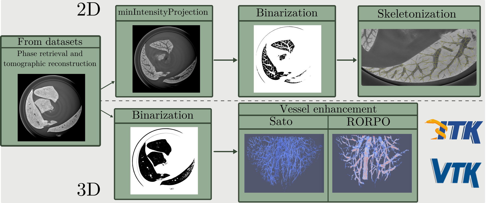
Extraction and reconstruction of mouse hepatic vascular network.
From phase contrast images, we were able to create the proof of concept about extraction of mouse hepatic vascular network [Work presented at the NEUBIAS conference in 2018]. All methodologies employed (see figure below) allowed us to assess the feasability of a complete pipeline from data acquisition, to analysis of them and finally, high level information extraction.
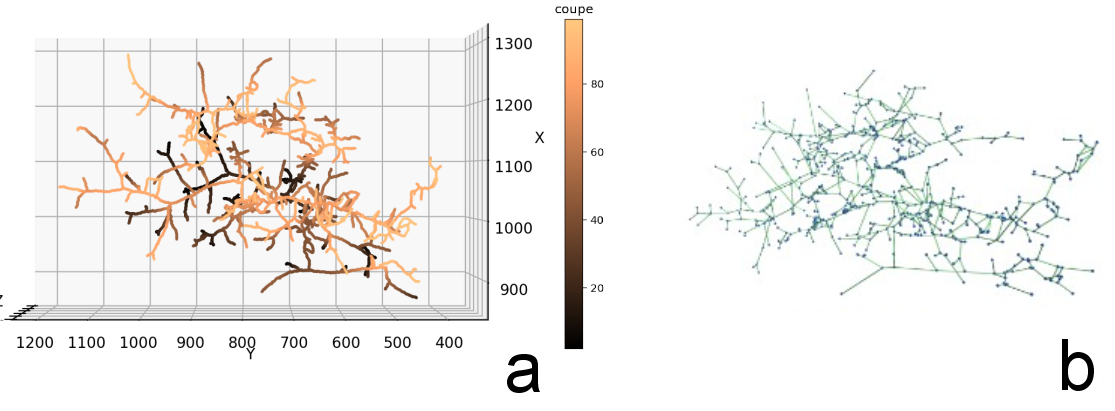
This topic consists of my main research activity and since i joined LORIA lab (ADAGIO team) i am even more convinced that we need to focus our effort on the nature of studied discrete objects (example below of the graph representation of a vascular network), refine them and make good use of the information bring by their geometrical properties.
Two work axis are now clearly my priority: first, improve the accuracy of the segmentation methods and tools, second, develop methodologies to tackle the discontinuities problem always present in this kind of network application.
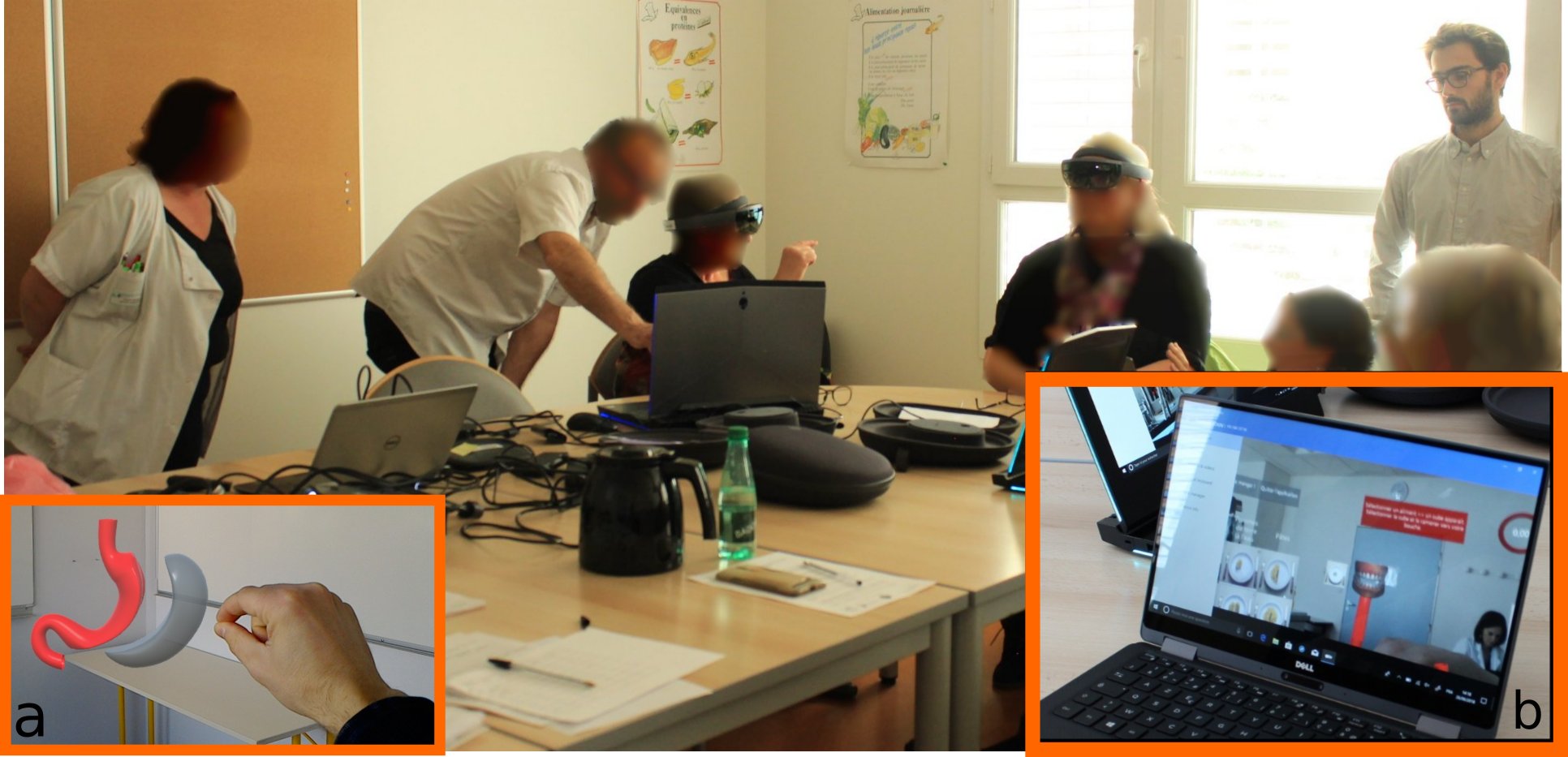
Mixed reality impact in a clinical context (2016-2020)
In the scope of athe AVACM project (CNRS PEPS INSIS), we were interested in measuring the impact of mixed reality in a medical context application. This project was designed to bring a specific type of mixed reality equipment (Microsoft HoloLens) towards patient to assess its acceptability and benefit in a clinical context.
This study was performed over 30 patients and showed that this kind of technology can be used in a medical context and present a good acceptability among the patient sample who participate. Moreover, a learning by the gesture can be performed and there improve the knowledge acquired by the patient [Barret-Grimault et al. (2019), Rositi et al. (2021), Appadoo et al. (2023)].
Participation in funded projects
| Name | Date (duration) | Project leader | Project proposal | Implication |
|---|---|---|---|---|
| PLBIO Plasma | Sept. 2022 (36 months) | S. Menecier (MCF UCA) | InCA | 17% |
| R-Vessel-X | Jan. 2019 (36 months) | A. Vacavant (PU UCA) | ANR | 30% |
| AVACM | Jan. 2017 (12 months) | C. Lohou (PU UCA) | PEPS INSIS | 50% |
-
PLBIO Plasma
Prostate cancer treatment by cold plasma application. -
R-Vessel-X
Robust vascular network extraction and understanding within hepatic biomedical images. -
AVACM
Augmented visual assistance for medical consultations.
Grants
-
NVIDIA Hardware grant ( August 2021, ad-hoc) - 2 GPUs AI dedicated.
Deep learning methods development for vascular structures segmentation. -
ESRF beamtime proposal ( October 2018, ad-hoc) - beamtime
X-ray phase contrast images acquisition of mouse livers.